
Acute Treatment of Osgood-Schlatter (Physical Therapy)

Osgood-Schlatter: treatment with physical therapy, diclofenac, and exercises.
Osgood-Schlatter syndrome is an osteochondrosis of the tibial tuberosity that mainly affects adolescents during periods of rapid growth and intense physical activity. It is characterized by pain, swelling, and tenderness at the tibial tuberosity. Below is a combined treatment plan with electrotherapy (iontophoresis with diclofenac), massage therapy, and kinesiotherapy, based on scientific evidence, for a patient with acute pain at the tibial plateaus due to Osgood-Schlatter.
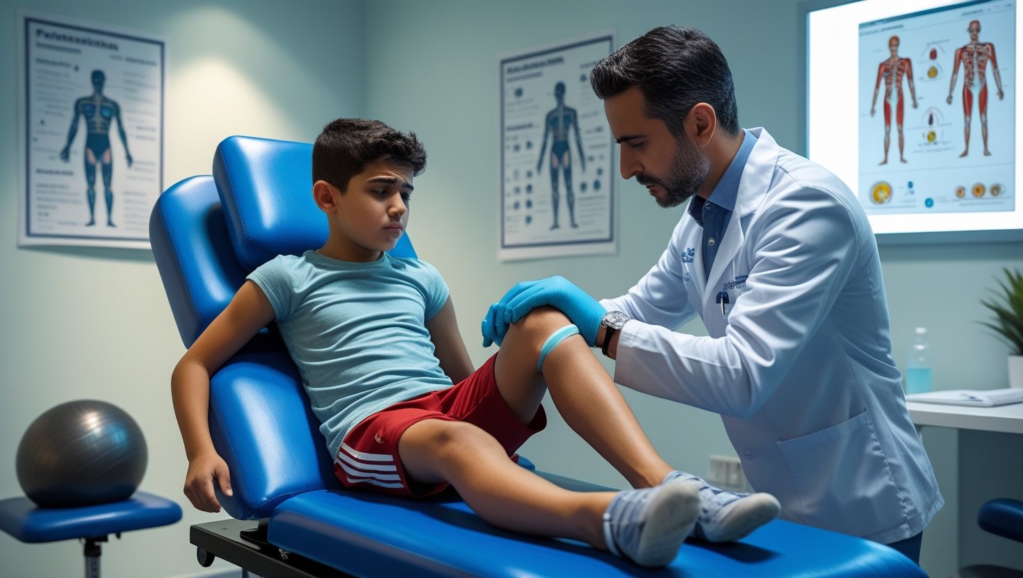
Treatment Goals:
- Reduce pain and inflammation.
- Improve function and mobility.
- Strengthen the surrounding musculature.
- Prevent recurrences.
- Promote a safe return to activity.
Treatment Plan
I. Acute Phase (Acute Pain)
A. Electrotherapy: Iontophoresis with Diclofenac
- Rationale: Iontophoresis is a technique that uses an electrical current to deliver ions of medicinal substances through the skin. Diclofenac is a nonsteroidal anti-inflammatory drug (NSAID) that can help reduce localized pain and inflammation.
- Scientific Evidence: Studies have demonstrated the effectiveness of iontophoresis with NSAIDs for managing localized musculoskeletal pain, offering an alternative to oral administration with fewer systemic side effects.
- Suggested Protocol:
- Medication: Diclofenac sodium in gel or solution (1-2% concentration).
- Active Electrode: Place the electrode with the diclofenac over the painful tibial tuberosity (negative polarity, since diclofenac is a negative ion).
- Dispersive Electrode: Place it at a distance of at least twice the diameter of the active electrode, preferably on the proximal part of the same thigh or on the calf.
- Current Intensity: Generally between 2-4 mA. The intensity should be comfortable for the patient, without causing burns or excessive irritation.
- Duration: 15-20 minutes per session.
- Frequency: 3-5 times per week, for 1-2 weeks, depending on the patient's response.
- Considerations: Assess skin integrity before and after each session. Avoid in patients with an allergy to diclofenac, a pacemaker, or metal implants in the area.
B. Massage Therapy
- Rationale: Massage therapy in this phase aims to reduce muscle spasm, improve local circulation, and promote relaxation of the tissues surrounding the tibial tuberosity.
- Suggested Protocol:
- Techniques: Gentle effleurage and petrissage massage around the quadriceps and hamstring musculature. Avoid direct pressure on the painful tibial tuberosity.
- Duration: 5-10 minutes per session.
- Frequency: Daily or every other day, depending on the patient's tolerance and pain intensity.
- Considerations: Perform with extreme gentleness. If the massage increases the pain, stop and reassess.
C. Kinesiotherapy (Initial Phase)
- Rationale: In the acute phase, kinesiotherapy focuses on maintaining mobility without aggravating the pain and on gentle muscle activation.
- Suggested Protocol:
- Gentle Passive/Assisted Mobilizations: Knee flexion-extension within the pain-free range of motion.
- Gentle Quadriceps Isometrics: Submaximal quadriceps contractions (20-30% of maximum voluntary contraction) with the knee extended, holding for 5-10 seconds, 10-15 repetitions. Perform several sets per day.
- Very Gentle Stretching: Passive or assisted stretching of the posterior chain (hamstrings and calves) and the quadriceps, without reaching the point of pain. Hold for 15-20 seconds, 2-3 repetitions. Perform 1-2 times a day.
- Patient Education:
- Relative rest from activities that cause pain (especially impact sports and jumping).
- Local cold application (ice pack) for 15-20 minutes, several times a day to reduce inflammation and pain.
- Use of knee braces with a patellar opening or a strap for the tibial tuberosity if deemed necessary to relieve pressure.
II. Recovery Phase (Pain Reduction, Functional Improvement)
Once the acute pain has decreased significantly.
A. Kinesiotherapy (Progression)
- Progressive Strengthening:
- Quadriceps: Knee extension with a resistance band or light weights (e.g., straight leg raises lying on your back, seated knee extension). Progress in load and repetitions.
- Hamstrings: Hamstring curls with a resistance band or body weight.
- Glutes: Glute bridges, Bulgarian split squats (modified according to tolerance), hip abduction.
- Core Muscles: Planks, bird-dog, etc. (important for overall stability and lower-limb biomechanics).
- Intensified Stretching:
- Quadriceps: Standing or prone quadriceps stretches.
- Hamstrings and Calves: Posterior chain stretches.
- Iliotibial Band: Specific stretches.
- Hold for 30 seconds, 3-5 repetitions, 2-3 times a day.
- Proprioceptive and Balance Exercises:
- Single-leg stance, balance board, exercises with eyes closed.
- Progression from stable to unstable surfaces.
- Motor Control and Coordination:
- Functional exercises that simulate sports movements but in a controlled way and without initial impact (e.g., mini squats, gentle lunges).
B. Activity Modification
- Gradual reintroduction of physical and sports activity.
- Avoid high-impact activities and jumping until the patient is completely symptom-free and has regained strength and control.
- Emphasize the importance of a proper warm-up and a cool-down after exercise.
General Considerations and Additional Evidence:
- Individualization: This plan is a guide. Every patient is unique, and the treatment must be adapted to their progress, pain tolerance, and response.
- Education: It is essential to educate the patient and their parents about the nature of Osgood-Schlatter, its benign course, and the importance of adherence to treatment and activity modification.
- Pain Management: In addition to iontophoresis, the use of ice after activity or exercises can be considered. In cases of very intense pain, and under medical supervision, oral analgesics may be considered.
- Shoes and Orthotics: Assess appropriate athletic footwear. Some patients may benefit from orthopedic insoles if there are underlying biomechanical problems.
- Slow Progression: The key to success in Osgood-Schlatter is a gradual progression of activity and exercises, avoiding premature overload of the tibial tuberosity.
- Scientific Evidence for Kinesiotherapy: The scientific literature strongly supports the importance of an individualized exercise program that includes strengthening of the quadriceps, hamstrings, and glutes, as well as stretching and proprioception, as the cornerstone of conservative treatment for Osgood-Schlatter. (Rathleff et al., 2014; Gholve et al., 2007).
- Prognosis: Osgood-Schlatter is typically self-limiting and resolves with the closure of the growth plates, although symptoms may persist for months or even years. Successful conservative treatment allows most adolescents to continue with their activities.
Treatment Duration:
Treatment time can vary considerably, from several weeks to several months, depending on the severity of symptoms, the patient's age, and adherence to the program. The goal is for the patient to return to their activities pain-free.
Key References (examples; consulting up-to-date literature is recommended):
- Rathleff, M. S., Roos, E. M., Olesen, J. L., & Jensen, M. B. (2014). High-volume, low-load training is effective in patients with patellofemoral pain: a randomised controlled trial. British Journal of Sports Medicine, 48(19), 1432-1436. (Although focused on patellofemoral pain, the strengthening principles are relevant).
- Gholve, P. A., Hosalkar, H. S., Hewlett, A., Gillogly, L. A., & Herman, M. J. (2007). Osgood Schlatter syndrome. Current Opinion in Pediatrics, 19(1), 44-50.
- Khan, K. M., & Scott, A. (2009). Mechanotherapy: how physical therapists' prescription of exercise promotes tissue repair. British Journal of Sports Medicine, 43(4), 247-251. (General loading principles for tissue healing).
- Specific articles on iontophoresis for localized musculoskeletal pain and the use of topical NSAIDs.
This plan should be supervised by a physical therapist with experience in pediatric musculoskeletal injuries.
Do these symptoms sound familiar? Book your evaluation.
Book via WhatsApp